
Researchers have developed a technique for creating nanoparticles that carry two different cancer-killing drugs into the body and deliver those drugs to separate parts of the cancer cell where they will be most effective. The technique was developed by researchers at North Carolina State University and the University of North Carolina at Chapel Hill.
“In testing on laboratory mice, our technique resulted in significant improvement in breast cancer tumor reduction as compared to conventional treatment techniques,” says Dr. Zhen Gu, senior author of a paper on the research and an assistant professor in the joint biomedical engineering program at NC State and UNC-Chapel Hill.
“Cancer cells can develop resistance to chemotherapy drugs, but are less likely to develop resistance when multiple drugs are delivered simultaneously,” Gu says. “However, different drugs target different parts of the cancer cell. For example, the protein drug TRAIL is most effective against the cell membrane, while doxorubicin (Dox) is most effective when delivered to the nucleus. We’ve come up with a sequential and site-specific delivery technique that first delivers TRAIL to cancer cell membranes and then penetrates the membrane to deliver Dox to the nucleus.”
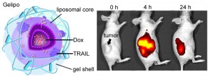
Image shows the structure of the nanoparticle (left), and how the nanoparticles home in on a tumor and shrink it (right). Image credit: North Carolina State University (Click image to enlarge)
Gu’s research team developed nanoparticles with an outer shell made of hyaluronic acid (HA) woven together with TRAIL. The HA interacts with receptors on cancer cell membranes, which “grab” the nanoparticle. Enzymes in the cancer cell environment break down the HA, releasing TRAIL onto the cell membrane and ultimately triggering cell death.
When the HA shell breaks down, it also reveals the core of the nanoparticle, which is made of Dox that is embedded with peptides that allow the core to penetrate into the cancer cell. The cancer cell encases the core in a protective bubble called an endosome, but the peptides on the core cause the endosome to begin breaking apart. This spills the Dox into the cell where it can penetrate the nucleus and trigger cell death.
“We designed this drug delivery vehicle using a ‘programmed’ strategy,” says Tianyue Jiang, a lead author in Dr. Gu’s lab. “Different drugs can be released at the right time in their right places,” adds Dr. Ran Mo, a postdoctoral researcher in Gu’s lab and the other lead author.
“This research is our first proof of concept, and we will continue to optimize the technique to make it even more efficient,” Gu says. “The early results are very promising, and we think this could be scaled up for large-scale manufacturing.”
The paper, “Gel–Liposome-Mediated Co-Delivery of Anticancer Membrane-Associated Proteins and Small-Molecule Drugs for Enhanced Therapeutic Efficacy,” is published online in Advanced Functional Materials. Co-authors of the paper are Adriano Bellotti, an undergraduate at NC State, and Dr. Jianping Zhou, a professor at China Pharmaceutical University.
*Source: North Carolina State University