
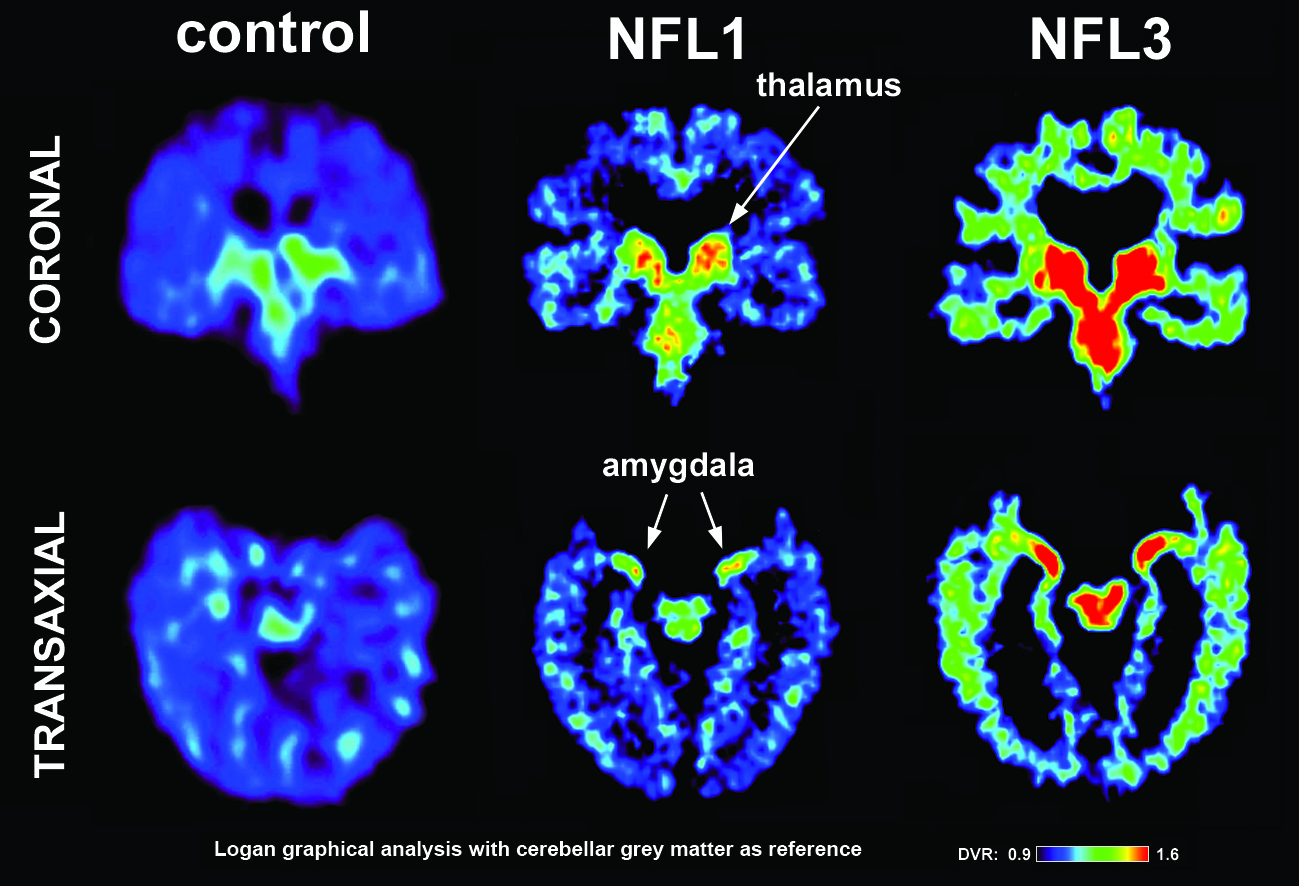
PET brain scans. Left image shows a normal brain scan; middle and right images show scans of pro football players from the study. Green and red colors demonstrate the higher level of tau protein found in the brain. Note the higher levels (more red and green) in the players’ scans. Scans of the players in the study reflect differing levels of tau protein and follow a pattern of progression similar to the tau deposits that have been observed at autopsy in CTE cases. Image courtesy of David Geffen School of Medicine at UCLA (Click image to enlarge)
(Visited 46 times, 1 visits today)