
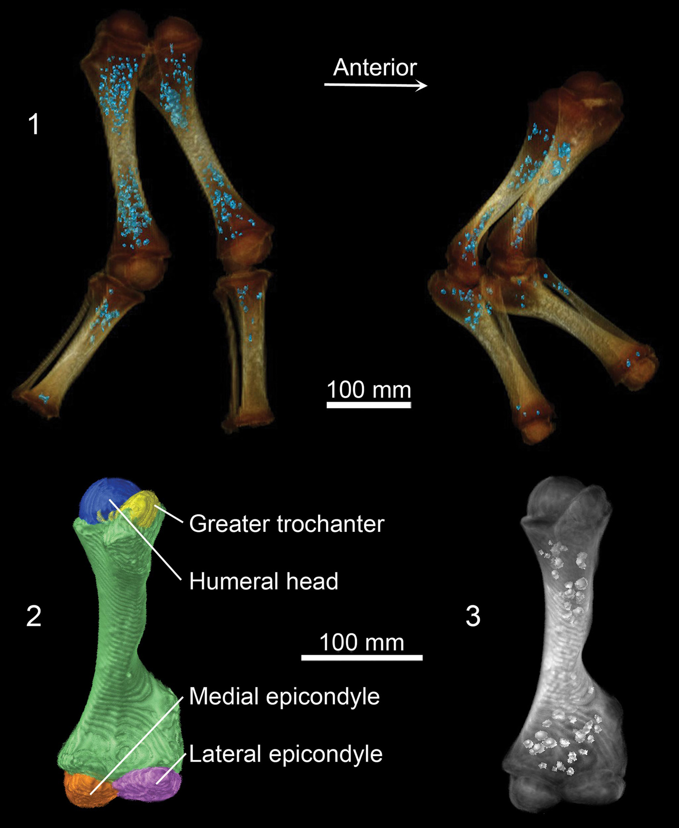
CT images showing Lyuba’s hind legs (top left) and front legs (top right). Bone shafts that are already well-hardened are white, and poorly mineralized ends of bones are brown. Blue dots within leg bones represent an iron phosphate mineral that follows the location of iron stores related to hemoglobin production while the calf was alive. Bottom images show Lyuba’s developing left humerus, or upper arm bone. Reflective spheroids on lower right image show location of iron phosphate mineral in this bone. Image credit: University of Michigan Museum of Paleontology (Click image to enlarge)
(Visited 23 times, 1 visits today)